Gangguan Jaras Penglihatan
Published on May 12, 2026
Risk Factors
Pituitary adenoma (chiasm), stroke in PCA territory (occipital), temporal lobe epilepsy surgery (Meyer's loop), multiple sclerosis (optic nerve/chiasm), craniopharyngioma (chiasm from above), trauma
Etiology
Compression, demyelination, ischemia, or neoplasm at any point along the pathway from retina to occipital cortex
Presentation
Painless, progressive visual field loss; may report bumping into objects on one side, difficulty driving, or reading trouble; acute onset suggests vascular etiology
Classic Exam
Confrontation visual field testing revealing a pattern-consistent defect; a relative afferent pupillary defect (RAPD/Marcus Gunn pupil) in optic nerve lesions; fundoscopic optic disc changes (pallor or papilledema)
Diagnostics
Formal perimetry (Humphrey or Goldmann) defining the field cut; MRI brain with gadolinium localizing the lesion; VEP (visual evoked potentials) for demyelinating optic neuritis
Management
Treat the underlying cause: surgical decompression for compressive masses, IV methylprednisolone for optic neuritis, stroke management for PCA infarcts
Keywords
"Bitemporal hemianopia," "homonymous hemianopia," "pie in the sky," "pie on the floor," "macular sparing," "RAPD," "tunnel vision"
01How the Visual Pathway Is Organized
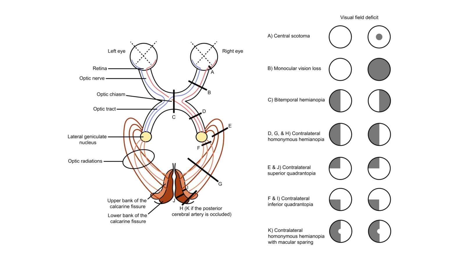
The visual pathway is a relay system that carries information from the retina to the primary visual cortex (V1, Brodmann area 17) in the occipital lobe. Understanding the fiber organization at each anatomical station is what allows you to localize a lesion based purely on the pattern of visual field loss.
Retinal organization and the rule of inversion. Light from the left visual field strikes the right half (nasal retina) of the left eye and the right half (temporal retina) of the right eye. This means that each eye captures both visual fields, but each hemifield is processed by a different retinal half. The nasal retinal fibers from each eye carry temporal visual field information, and the temporal retinal fibers carry nasal visual field information. This inversion is the foundation of every field defect pattern you will encounter.
The optic chiasm is the crossing point. At the chiasm, nasal fibers decussate (cross to the opposite side) while temporal fibers remain ipsilateral. After the chiasm, each optic tract therefore carries a complete representation of the contralateral visual field: the left optic tract carries the entire right visual field (from the left temporal retina + right nasal retina), and vice versa. This is why lesions posterior to the chiasm produce homonymous defects (affecting the same field in both eyes).
The lateral geniculate nucleus (LGN) is the thalamic relay station. It has six layers that maintain retinotopic organization and separate input from each eye (layers 2, 3, 5 from the ipsilateral eye; layers 1, 4, 6 from the contralateral eye). The LGN sends fibers posteriorly as the optic radiations, which split into two anatomically distinct bundles.
Meyer's loop (the temporal radiation) carries fibers representing the superior visual field. These fibers loop anteriorly and inferiorly through the temporal lobe before turning posteriorly to reach the inferior bank of the calcarine sulcus (lingual gyrus). Because of this detour through the temporal lobe, temporal lobe surgery or pathology selectively damages superior field fibers, producing a contralateral superior quadrantanopia ("pie in the sky").
The parietal radiation carries fibers representing the inferior visual field. These fibers travel more directly through the parietal lobe to reach the superior bank of the calcarine sulcus (cuneus). Parietal lesions therefore produce a contralateral inferior quadrantanopia ("pie on the floor").
Macular sparing is the hallmark of occipital cortex lesions. The macula has dual blood supply from both the posterior cerebral artery (PCA) and the middle cerebral artery (MCA), and it occupies a disproportionately large cortical representation at the occipital pole. When a PCA stroke damages the visual cortex, the macular representation is often spared because collateral flow from the MCA preserves it. This is why an occipital lesion produces a homonymous hemianopia with a preserved central island of vision, distinguishing it from an optic tract lesion that produces a complete homonymous hemianopia without macular sparing.
02Classification of Lesions and Their Visual Field Defects
Optic nerve
(pre-chiasmal)
Visual Field Defect
Ipsilateral monocular vision loss
Pattern Name
Monocular blindness
Key Feature
Only ONE eye affected; RAPD present on the affected side
Optic chiasm
(central)
Visual Field Defect
Loss of both temporal fields
Pattern Name
Bitemporal hemianopia
Key Feature
"Tunnel vision"; heteronymous (opposite halves in each eye)
Optic tract
(post-chiasmal)
Visual Field Defect
Loss of the contralateral visual field in both eyes
Pattern Name
Contralateral homonymous hemianopia
Key Feature
Incongruent (unequal field loss between the two eyes); RAPD on contralateral side
Temporal radiation
(Meyer's loop)
Visual Field Defect
Loss of contralateral superior quadrant
Pattern Name
Contralateral superior quadrantanopia
Key Feature
"Pie in the sky"; associated with temporal lobe lesions
Parietal radiation
Visual Field Defect
Loss of contralateral inferior quadrant
Pattern Name
Contralateral inferior quadrantanopia
Key Feature
"Pie on the floor"; associated with parietal lobe lesions
Occipital cortex (V1)
Visual Field Defect
Loss of contralateral visual field with central sparing
Pattern Name
Contralateral homonymous hemianopia with macular sparing
Key Feature
Highly congruent; macular sparing due to dual blood supply
Lesion Site | Visual Field Defect | Pattern Name | Key Feature |
|---|---|---|---|
Optic nerve | Ipsilateral monocular vision loss | Monocular blindness | Only ONE eye affected; RAPD present on the affected side |
Optic chiasm | Loss of both temporal fields | Bitemporal hemianopia | "Tunnel vision"; heteronymous (opposite halves in each eye) |
Optic tract | Loss of the contralateral visual field in both eyes | Contralateral homonymous hemianopia | Incongruent (unequal field loss between the two eyes); RAPD on contralateral side |
Temporal radiation | Loss of contralateral superior quadrant | Contralateral superior quadrantanopia | "Pie in the sky"; associated with temporal lobe lesions |
Parietal radiation | Loss of contralateral inferior quadrant | Contralateral inferior quadrantanopia | "Pie on the floor"; associated with parietal lobe lesions |
Occipital cortex (V1) | Loss of contralateral visual field with central sparing | Contralateral homonymous hemianopia with macular sparing | Highly congruent; macular sparing due to dual blood supply |
Congruence is a useful localizing concept. Lesions closer to the cortex produce more congruent defects (the field loss is nearly identical in both eyes), while lesions closer to the chiasm produce more incongruent defects. An optic tract lesion causes an incongruent homonymous hemianopia; an occipital cortex lesion causes a highly congruent one. This distinction can appear as the discriminating detail in a vignette.
Special Chiasmal Variants
Central chiasmal compression (pituitary adenoma pushing up)
Mechanism
Crossing nasal fibers compressed
Field Defect
Bitemporal hemianopia
Lateral chiasmal compression (internal carotid aneurysm)
Mechanism
Ipsilateral uncrossed temporal fibers compressed
Field Defect
Ipsilateral nasal hemianopia (rare, but testable)
Anterior junction syndrome
Mechanism
Lesion at junction of optic nerve and chiasm; damages ipsilateral nerve + contralateral inferior nasal fibers (Wilbrand's knee)
Field Defect
Ipsilateral monocular blindness + contralateral superior temporal quadrant defect (junctional scotoma)
Variant | Mechanism | Field Defect |
|---|---|---|
Central chiasmal compression (pituitary adenoma pushing up) | Crossing nasal fibers compressed | Bitemporal hemianopia |
Lateral chiasmal compression (internal carotid aneurysm) | Ipsilateral uncrossed temporal fibers compressed | Ipsilateral nasal hemianopia (rare, but testable) |
Anterior junction syndrome | Lesion at junction of optic nerve and chiasm; damages ipsilateral nerve + contralateral inferior nasal fibers (Wilbrand's knee) | Ipsilateral monocular blindness + contralateral superior temporal quadrant defect (junctional scotoma) |
03Diagnostic Workup
Confrontation visual field testing
Role
Best initial bedside test
What It Shows
Identifies gross field cuts; each quadrant tested separately
Formal automated perimetry (Humphrey)
Role
Best initial quantitative test
What It Shows
Maps precise field defect boundaries; standard for monitoring progression
Goldmann perimetry
Role
Alternative for patients who cannot perform automated testing
What It Shows
Kinetic perimetry; useful in neurological defects and malingering workup
MRI brain with gadolinium
Role
Most accurate test for localizing the lesion
What It Shows
Shows masses (pituitary adenoma, craniopharyngioma, meningioma), demyelination plaques, or infarcts along the pathway
Visual evoked potentials (VEP)
Role
Best test for optic nerve demyelination
What It Shows
Prolonged P100 latency confirms optic neuritis even after clinical recovery
Optical coherence tomography (OCT)
Role
Adjunct for optic nerve disease
What It Shows
Measures retinal nerve fiber layer thickness; thinning confirms axonal loss
Pupillary light reflex
Role
Bedside test for afferent pathway integrity
What It Shows
RAPD (swinging flashlight test) localizes to optic nerve or extensive retinal disease
Test | Role | What It Shows |
|---|---|---|
Confrontation visual field testing | Best initial bedside test | Identifies gross field cuts; each quadrant tested separately |
Formal automated perimetry (Humphrey) | Best initial quantitative test | Maps precise field defect boundaries; standard for monitoring progression |
Goldmann perimetry | Alternative for patients who cannot perform automated testing | Kinetic perimetry; useful in neurological defects and malingering workup |
MRI brain with gadolinium | Most accurate test for localizing the lesion | Shows masses (pituitary adenoma, craniopharyngioma, meningioma), demyelination plaques, or infarcts along the pathway |
Visual evoked potentials (VEP) | Best test for optic nerve demyelination | Prolonged P100 latency confirms optic neuritis even after clinical recovery |
Optical coherence tomography (OCT) | Adjunct for optic nerve disease | Measures retinal nerve fiber layer thickness; thinning confirms axonal loss |
Pupillary light reflex | Bedside test for afferent pathway integrity | RAPD (swinging flashlight test) localizes to optic nerve or extensive retinal disease |
Workup logic. When a patient presents with visual field loss, the first step is confrontation testing to identify the pattern. This alone tells you the anatomical level. Once you know the pattern, you order MRI brain with contrast to identify the structural cause. The sequence is always: define the field defect first, then image.
The swinging flashlight test is a high-yield bedside maneuver. A relative afferent pupillary defect (RAPD) is present when the affected eye's pupil paradoxically dilates when light swings from the normal eye to the affected eye. This indicates optic nerve disease (pre-chiasmal) or an extensive retinal lesion. An RAPD is not seen in chiasmal or retrochiasmal lesions because both eyes' afferent input is affected symmetrically in those cases. One important exception: an optic tract lesion can produce a contralateral RAPD because the tract carries more crossed (nasal) fibers than uncrossed (temporal) fibers, creating asymmetry.
When to order VEP. If the clinical picture suggests optic neuritis (young woman with painful monocular vision loss that recovers over weeks), VEP is the test that confirms prior demyelination even after vision has returned to baseline. The P100 latency remains prolonged indefinitely, serving as an objective record of a past episode.
04Management and Treatment
Pituitary adenoma (bitemporal hemianopia)
Acute Management
Urgent surgical evaluation; if apoplexy, emergent decompression
Long-Term Management
Transsphenoidal resection; hormonal replacement as needed; cabergoline/bromocriptine if prolactinoma
Optic neuritis (monocular vision loss)
Acute Management
IV methylprednisolone 1 g/day for 3-5 days, then oral prednisone taper over 11 days
Long-Term Management
Evaluate for MS with MRI brain/spine; consider disease-modifying therapy if demyelinating lesions present
PCA stroke (homonymous hemianopia with macular sparing)
Acute Management
Acute stroke protocol: thrombolytics if within window, thrombectomy if large vessel occlusion
Long-Term Management
Secondary stroke prevention: antiplatelets, statins, blood pressure control, rehabilitation
Temporal lobe tumor/surgery (superior quadrantanopia)
Acute Management
Surgical planning to minimize Meyer's loop damage; preoperative tractography
Long-Term Management
Post-surgical monitoring; neuro-rehabilitation if field cut is disabling
Craniopharyngioma (bitemporal hemianopia)
Acute Management
Surgical resection (transcranial or transsphenoidal depending on extension)
Long-Term Management
Lifelong pituitary hormone monitoring and replacement; risk of recurrence
Compressive meningioma
Acute Management
Surgical excision
Long-Term Management
Serial imaging; radiation therapy if subtotal resection
Etiology | Acute Management | Long-Term Management |
|---|---|---|
Pituitary adenoma (bitemporal hemianopia) | Urgent surgical evaluation; if apoplexy, emergent decompression | Transsphenoidal resection; hormonal replacement as needed; cabergoline/bromocriptine if prolactinoma |
Optic neuritis (monocular vision loss) | IV methylprednisolone 1 g/day for 3-5 days, then oral prednisone taper over 11 days | Evaluate for MS with MRI brain/spine; consider disease-modifying therapy if demyelinating lesions present |
PCA stroke (homonymous hemianopia with macular sparing) | Acute stroke protocol: thrombolytics if within window, thrombectomy if large vessel occlusion | Secondary stroke prevention: antiplatelets, statins, blood pressure control, rehabilitation |
Temporal lobe tumor/surgery (superior quadrantanopia) | Surgical planning to minimize Meyer's loop damage; preoperative tractography | Post-surgical monitoring; neuro-rehabilitation if field cut is disabling |
Craniopharyngioma (bitemporal hemianopia) | Surgical resection (transcranial or transsphenoidal depending on extension) | Lifelong pituitary hormone monitoring and replacement; risk of recurrence |
Compressive meningioma | Surgical excision | Serial imaging; radiation therapy if subtotal resection |
Critical management pearl for optic neuritis: The Optic Neuritis Treatment Trial (ONTT) demonstrated that IV methylprednisolone speeds visual recovery and reduces the short-term risk of MS development, but oral prednisone alone (without preceding IV therapy) actually increases recurrence risk. Never start with oral steroids alone for optic neuritis. The correct sequence is IV first, then oral taper.
Pituitary apoplexy is a surgical emergency. When a patient with a known pituitary adenoma develops sudden-onset bitemporal hemianopia, severe headache, and ophthalmoplegia (compression of cranial nerves III, IV, VI in the cavernous sinus), the next best step is emergent surgical decompression along with IV corticosteroids for adrenal crisis.
05Differential Diagnosis and Distractors
Retinal detachment vs. optic nerve lesion
Why It Looks Similar
Both cause monocular vision loss
Key Discriminator
Retinal detachment: "curtain falling" over vision, no RAPD unless extensive; fundoscopy shows retinal elevation. Optic nerve lesion: RAPD present, disc pallor or edema on fundoscopy
Open-angle glaucoma vs. chiasmal compression
Why It Looks Similar
Both can cause bilateral visual field loss progressing inward
Key Discriminator
Glaucoma: elevated IOP, peripheral nasal step or arcuate scotoma respecting the horizontal meridian. Chiasmal lesion: defect respects the vertical meridian (temporal fields lost bilaterally)
Optic tract lesion vs. occipital cortex stroke
Why It Looks Similar
Both produce contralateral homonymous hemianopia
Key Discriminator
Tract: incongruent, may have contralateral RAPD, optic disc pallor (bow-tie atrophy). Occipital: highly congruent, macular sparing, no RAPD, no disc changes
Anterior ischemic optic neuropathy (AION) vs. optic neuritis
Why It Looks Similar
Both cause acute monocular vision loss with RAPD
Key Discriminator
AION: painless, older patient (>50), "disc at risk" (small cup-to-disc ratio), altitudinal field defect, pallid disc edema. Optic neuritis: painful (worse with eye movement), younger patient (20-40), central scotoma
Functional (non-organic) visual loss vs. true field defect
Why It Looks Similar
Patient reports complete hemianopia but exam findings are inconsistent
Key Discriminator
Functional: field defect that does not expand with distance testing (tubular field), OKN (optokinetic nystagmus) response preserved toward the "blind" field, normal pupillary responses and VEP
Migraine with aura vs. occipital infarct
Why It Looks Similar
Both produce transient homonymous visual disturbance
Key Discriminator
Migraine: scintillating scotoma lasting 15-30 minutes that marches across the field, followed by headache, fully reversible. Stroke: sudden onset, persistent, no scintillation
Differential | Why It Looks Similar | Key Discriminator |
|---|---|---|
Retinal detachment vs. optic nerve lesion | Both cause monocular vision loss | Retinal detachment: "curtain falling" over vision, no RAPD unless extensive; fundoscopy shows retinal elevation. Optic nerve lesion: RAPD present, disc pallor or edema on fundoscopy |
Open-angle glaucoma vs. chiasmal compression | Both can cause bilateral visual field loss progressing inward | Glaucoma: elevated IOP, peripheral nasal step or arcuate scotoma respecting the horizontal meridian. Chiasmal lesion: defect respects the vertical meridian (temporal fields lost bilaterally) |
Optic tract lesion vs. occipital cortex stroke | Both produce contralateral homonymous hemianopia | Tract: incongruent, may have contralateral RAPD, optic disc pallor (bow-tie atrophy). Occipital: highly congruent, macular sparing, no RAPD, no disc changes |
Anterior ischemic optic neuropathy (AION) vs. optic neuritis | Both cause acute monocular vision loss with RAPD | AION: painless, older patient (>50), "disc at risk" (small cup-to-disc ratio), altitudinal field defect, pallid disc edema. Optic neuritis: painful (worse with eye movement), younger patient (20-40), central scotoma |
Functional (non-organic) visual loss vs. true field defect | Patient reports complete hemianopia but exam findings are inconsistent | Functional: field defect that does not expand with distance testing (tubular field), OKN (optokinetic nystagmus) response preserved toward the "blind" field, normal pupillary responses and VEP |
Migraine with aura vs. occipital infarct | Both produce transient homonymous visual disturbance | Migraine: scintillating scotoma lasting 15-30 minutes that marches across the field, followed by headache, fully reversible. Stroke: sudden onset, persistent, no scintillation |
06Traps and High-Yield Pearls
The single most tested concept in visual pathway questions is your ability to match a field defect pattern to a lesion site. The vignette will describe a patient's visual complaint, and you must reason backward: is this monocular or binocular? Does it respect the vertical or horizontal meridian? Is it homonymous or heteronymous? Is there macular sparing?
A common trap is confusing bitemporal hemianopia (chiasm) with binasal hemianopia. Binasal hemianopia would require bilateral compression of the uncrossed temporal fibers from both sides, which is anatomically extremely rare and almost never the answer. If a vignette describes bilateral temporal field loss, the answer is chiasmal compression, almost always from a pituitary adenoma growing upward.
Another frequently tested mistake is failing to distinguish optic tract from occipital cortex lesions. Both produce contralateral homonymous hemianopia. The discriminators are: the tract lesion is incongruent with a possible RAPD, while the cortical lesion is highly congruent with macular sparing and no pupillary abnormality.
The mnemonic "pie in the sky, pie on the floor" is high-yield: temporal lobe (Meyer's loop) damage loses the superior quadrant (pie in the sky), parietal lobe damage loses the inferior quadrant (pie on the floor). When a vignette describes a patient who underwent temporal lobectomy for epilepsy and now has a new visual field deficit, the answer is contralateral superior quadrantanopia.
Finally, remember that RAPD is a pre-chiasmal finding. If a vignette gives you a patient with a homonymous hemianopia and an RAPD, think optic tract, not occipital cortex. The presence or absence of an RAPD is often the single detail that localizes the lesion for you.