Interrupted Aortic Arch (IAA)
Published on May 22, 2026
Risk Factors
DiGeorge syndrome (22q11.2 microdeletion), maternal diabetes, fetal alcohol exposure; strong association with chromosome 22q11 deletion (up to 50% of Type B cases)
Etiology
Congenital absence of a segment of the aortic arch, resulting in complete luminal discontinuity between the ascending and descending aorta; ductal-dependent systemic circulation
Presentation
Neonate presents with acute cardiovascular collapse, poor feeding, tachypnea, and severe metabolic acidosis within the first days of life as the ductus arteriosus closes
Classic Exam
Differential cyanosis (lower body cyanotic, upper body pink in Type A), absent or diminished femoral pulses, blood pressure discrepancy between upper and lower extremities, signs of congestive heart failure
Diagnostics
Echocardiography showing complete discontinuity of the aortic arch; chest X-ray with cardiomegaly and pulmonary edema; severe lactic acidosis on arterial blood gas
Management
Immediate prostaglandin E1 (PGE1) infusion to maintain ductal patency, followed by surgical repair (direct anastomosis or interposition graft)
Keywords
"Ductal-dependent lesion," "cardiovascular collapse in a neonate," "absent femoral pulses," "DiGeorge syndrome," "22q11.2 deletion," "prostaglandin E1"
01Pathophysiology
Interrupted aortic arch (IAA) is a congenital cardiac defect characterized by complete anatomic discontinuity of the aortic arch. Unlike coarctation, where a narrowing exists but some luminal continuity remains, IAA involves a full gap in the arch. This means the ascending aorta and the descending aorta are physically separated, and blood cannot flow from one to the other through the arch itself.
During fetal life, this defect is well tolerated because the ductus arteriosus serves as the bridge. Blood ejected from the right ventricle into the pulmonary artery crosses the patent ductus arteriosus (PDA) and supplies the descending aorta, perfusing the lower body, kidneys, and gut. The fetus remains hemodynamically stable because ductal flow compensates entirely for the absent arch segment.
The crisis begins after birth when the ductus arteriosus undergoes physiologic closure, typically within the first 24 to 72 hours of life. As the duct constricts, systemic perfusion to the lower body is abruptly cut off. The kidneys become ischemic, the gut is underperfused, and the neonate develops progressive lactic acidosis from anaerobic metabolism in the lower body. Simultaneously, the left ventricle faces an enormous afterload because all cardiac output must now be directed through whatever proximal arch exists, leading to rapid-onset congestive heart failure with pulmonary edema.
Nearly all cases of IAA are associated with a ventricular septal defect (VSD), which is present in over 95% of patients. The VSD allows some degree of intracardiac mixing but does not prevent the hemodynamic catastrophe that occurs once the ductus closes. The VSD is an important associated finding because it influences the surgical approach.
The strong link to 22q11.2 microdeletion (DiGeorge syndrome) is a high-yield association. This chromosomal abnormality affects neural crest cell migration, which is critical for the development of the aortic arch derivatives, the thymus, and the parathyroid glands. A neonate with IAA who also has hypocalcemia (from absent parathyroids), T-cell lymphopenia (from thymic hypoplasia), and characteristic facial features should immediately prompt genetic testing for 22q11.2 deletion.
02Classification and Clinical Manifestation
Celoria-Patton Classification
A
Site of Interruption
Distal to the left subclavian artery (between left subclavian and descending aorta)
Frequency
~30-40%
Key Association
Resembles a severe coarctation location; less associated with DiGeorge
B
Site of Interruption
Between the left common carotid artery and the left subclavian artery
Frequency
~50-60% (most common)
Key Association
Strongest association with 22q11.2 deletion and DiGeorge syndrome
C
Site of Interruption
Between the innominate (brachiocephalic) artery and the left common carotid artery
Frequency
~5% (rare)
Key Association
Least common variant
Type | Site of Interruption | Frequency | Key Association |
|---|---|---|---|
A | Distal to the left subclavian artery (between left subclavian and descending aorta) | ~30-40% | Resembles a severe coarctation location; less associated with DiGeorge |
B | Between the left common carotid artery and the left subclavian artery | ~50-60% (most common) | Strongest association with 22q11.2 deletion and DiGeorge syndrome |
C | Between the innominate (brachiocephalic) artery and the left common carotid artery | ~5% (rare) | Least common variant |
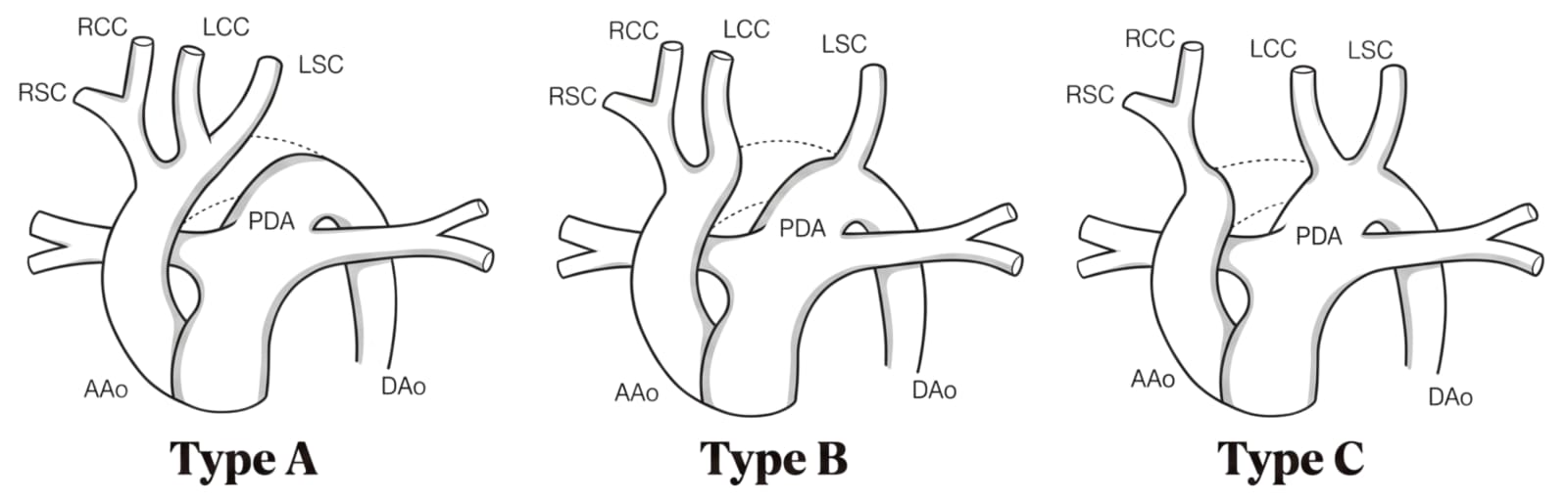
The hallmark physical finding is differential pulse quality and blood pressure between the upper and lower extremities.
In Type A, the right arm, left arm, and head all receive flow from the ascending aorta, while the lower body depends on ductal flow.
In Type B, the left subclavian artery arises distal to the interruption, so the left arm may also have diminished pulses, creating an asymmetry even between the two upper extremities. This is a testable nuance that distinguishes Type B from Type A and from coarctation.
By Timing
Timing | Clinical Features |
|---|---|
First 24-48 hours (ductus open) | Neonate may appear deceptively well; mild tachypnea; may have a murmur from the associated VSD; pulses may initially be palpable if the ductus is widely patent |
48-72 hours (ductus closing) | Acute deterioration with tachypnea, poor feeding, lethargy, mottled or gray skin, weak or absent femoral pulses, oliguria or anuria |
After ductal closure | Cardiovascular collapse, profound metabolic acidosis (, ), multiorgan failure (renal, hepatic, intestinal ischemia), cardiogenic shock |
03Diagnostic Workup
Echocardiography
Role
Best initial and confirmatory test
Expected Findings
Complete discontinuity of the aortic arch; PDA providing descending aortic flow; associated VSD; assessment of arch anatomy and branch vessel origins
Chest X-ray
Role
Supportive
Expected Findings
Cardiomegaly, pulmonary vascular congestion/edema, absent or abnormal aortic knob silhouette
Arterial blood gas
Role
Assess severity
Expected Findings
Severe metabolic acidosis with elevated lactate; low in post-ductal samples
Four-limb blood pressure
Role
Screening at bedside
Expected Findings
Systolic pressure gradient between upper and lower extremities >10 mmHg; in Type B, discrepancy between right and left arms
CT angiography
Role
Anatomic delineation (if echo is inconclusive)
Expected Findings
Precise visualization of the arch anatomy, gap location, and branch vessel relationships for surgical planning
Genetic testing (FISH for 22q11.2)
Role
Rule out DiGeorge
Expected Findings
Indicated in all cases of IAA, particularly Type B
Serum calcium, T-cell subsets
Role
Screen for DiGeorge features
Expected Findings
Hypocalcemia from hypoparathyroidism; lymphopenia with reduced CD3+ T-cells
Test | Role | Expected Findings |
|---|---|---|
Echocardiography | Best initial and confirmatory test | Complete discontinuity of the aortic arch; PDA providing descending aortic flow; associated VSD; assessment of arch anatomy and branch vessel origins |
Chest X-ray | Supportive | Cardiomegaly, pulmonary vascular congestion/edema, absent or abnormal aortic knob silhouette |
Arterial blood gas | Assess severity | Severe metabolic acidosis with elevated lactate; low in post-ductal samples |
Four-limb blood pressure | Screening at bedside | Systolic pressure gradient between upper and lower extremities >10 mmHg; in Type B, discrepancy between right and left arms |
CT angiography | Anatomic delineation (if echo is inconclusive) | Precise visualization of the arch anatomy, gap location, and branch vessel relationships for surgical planning |
Genetic testing (FISH for 22q11.2) | Rule out DiGeorge | Indicated in all cases of IAA, particularly Type B |
Serum calcium, T-cell subsets | Screen for DiGeorge features | Hypocalcemia from hypoparathyroidism; lymphopenia with reduced CD3+ T-cells |
Echocardiography is both the best initial test and often the definitive diagnostic study. In an acutely ill neonate with absent femoral pulses and metabolic acidosis, bedside echocardiography can rapidly identify the arch discontinuity, demonstrate retrograde flow through the PDA into the descending aorta, and characterize the associated VSD. This is the study that should be ordered first.
The four-limb blood pressure and pulse oximetry assessment is a bedside screening maneuver. A saturation difference between the right hand (pre-ductal) and the feet (post-ductal) greater than 3% suggests ductal-dependent lower body perfusion. This can also be picked up on routine newborn pulse oximetry screening, which is why critical congenital heart disease screening programs exist.
CT angiography or cardiac MRI is reserved for cases where echocardiography does not provide adequate anatomic detail or when the surgical team needs precise three-dimensional reconstruction of the arch anatomy. These are not first-line tests in the acute setting.
Genetic evaluation should not be delayed. Given that up to 50% of Type B IAA patients carry the 22q11.2 deletion, fluorescence in situ hybridization (FISH) or chromosomal microarray should be sent early. The presence of DiGeorge syndrome has implications for surgical planning (irradiated blood products to prevent graft-versus-host disease due to T-cell immunodeficiency), calcium management, and long-term follow-up.
04Management and Treatment
Immediate stabilization
Intervention
Prostaglandin E1 (PGE1) infusion
Details
Starting dose: IV; titrate down to once the ductus reopens; goal is to restore ductal patency and lower body perfusion
Supportive care
Intervention
Mechanical ventilation, inotropes, correction of acidosis
Details
Intubation (PGE1 causes apnea); dopamine or milrinone for cardiac support; sodium bicarbonate if ; correct hypocalcemia if DiGeorge is present
Definitive treatment
Intervention
Surgical repair
Details
Direct anastomosis of the aortic arch segments (primary repair) with simultaneous VSD closure; performed under cardiopulmonary bypass with deep hypothermic circulatory arrest; typically done within the first week of life
Postoperative
Intervention
Long-term surveillance
Details
Serial echocardiography to monitor for re-stenosis at the repair site; blood pressure monitoring; genetic counseling and immunologic follow-up if 22q11.2 deletion confirmed
Phase | Intervention | Details |
|---|---|---|
Immediate stabilization | Prostaglandin E1 (PGE1) infusion | Starting dose: IV; titrate down to once the ductus reopens; goal is to restore ductal patency and lower body perfusion |
Supportive care | Mechanical ventilation, inotropes, correction of acidosis | Intubation (PGE1 causes apnea); dopamine or milrinone for cardiac support; sodium bicarbonate if ; correct hypocalcemia if DiGeorge is present |
Definitive treatment | Surgical repair | Direct anastomosis of the aortic arch segments (primary repair) with simultaneous VSD closure; performed under cardiopulmonary bypass with deep hypothermic circulatory arrest; typically done within the first week of life |
Postoperative | Long-term surveillance | Serial echocardiography to monitor for re-stenosis at the repair site; blood pressure monitoring; genetic counseling and immunologic follow-up if 22q11.2 deletion confirmed |
PGE1 is the single most critical initial intervention. The moment IAA is suspected, PGE1 must be started without waiting for echocardiographic confirmation. The drug relaxes the smooth muscle of the ductus arteriosus, reopening it and restoring blood flow to the descending aorta. Clinically, you will see the femoral pulses return, urine output improve, and the lactate begin to clear. The most important side effect to monitor is apnea, which occurs in 10-12% of neonates on PGE1 and necessitates elective intubation in many centers before transport.
Metabolic acidosis should be corrected with careful fluid resuscitation and, if severe, sodium bicarbonate. Hypocalcemia must be checked and corrected promptly, particularly because unrecognized DiGeorge syndrome can cause refractory hypocalcemia that worsens cardiac contractility and contributes to hemodynamic instability.
Surgical repair is performed as a single-stage procedure in most centers: the arch is reconstructed by direct end-to-end anastomosis (or with a homograft patch if the gap is too wide), and the VSD is closed simultaneously through a median sternotomy. The surgery is typically performed within the first 1 to 2 weeks of life, once the neonate is stabilized.
Contraindication to keep in mind: If a neonate with IAA and confirmed DiGeorge syndrome requires blood transfusion (perioperatively or otherwise), all blood products must be irradiated and CMV-negative to prevent transfusion-associated graft-versus-host disease, given the underlying T-cell immunodeficiency.
Long-term complications include re-coarctation at the surgical repair site (the most common late complication, occurring in up to 25% of patients), subaortic stenosis from the VSD patch or left ventricular outflow tract narrowing, and aortic valve disease. These patients require lifelong cardiology follow-up.
05Differential Diagnosis and Distractors
Coarctation of the aorta
Why It Looks Similar
Both present with upper-lower extremity blood pressure gradient, diminished femoral pulses, and can be ductal-dependent in neonates
Key Discriminator
Coarctation has a narrowing but not complete discontinuity; echo shows a discrete shelf or ridge with some antegrade flow through the arch; IAA shows no flow across the gap
Hypoplastic left heart syndrome (HLHS)
Why It Looks Similar
Both are ductal-dependent systemic circulation lesions presenting with shock in the first days of life
Key Discriminator
HLHS has an underdeveloped left ventricle and aortic/mitral valve atresia or stenosis; the entire systemic output comes from the right ventricle through the PDA; IAA typically has two functioning ventricles with a VSD
Critical aortic stenosis
Why It Looks Similar
Both can present with low cardiac output and shock in neonates
Key Discriminator
Critical aortic stenosis shows a thickened, immobile aortic valve with high-velocity jet on echo; the arch itself is intact and continuous
Neonatal sepsis
Why It Looks Similar
Both present with poor feeding, tachypnea, metabolic acidosis, and hemodynamic collapse in a neonate
Key Discriminator
Sepsis will not show a blood pressure or pulse discrepancy between upper and lower extremities; echocardiography is structurally normal; blood cultures are positive
Truncus arteriosus
Why It Looks Similar
Also strongly associated with 22q11.2 deletion and DiGeorge syndrome; presents early in life with heart failure
Key Discriminator
Truncus has a single arterial trunk arising from the heart supplying both systemic and pulmonary circulations; the arch, while sometimes abnormal, is not interrupted in the classic sense (though IAA can coexist with truncus)
Diagnosis | Why It Looks Similar | Key Discriminator |
|---|---|---|
Coarctation of the aorta | Both present with upper-lower extremity blood pressure gradient, diminished femoral pulses, and can be ductal-dependent in neonates | Coarctation has a narrowing but not complete discontinuity; echo shows a discrete shelf or ridge with some antegrade flow through the arch; IAA shows no flow across the gap |
Hypoplastic left heart syndrome (HLHS) | Both are ductal-dependent systemic circulation lesions presenting with shock in the first days of life | HLHS has an underdeveloped left ventricle and aortic/mitral valve atresia or stenosis; the entire systemic output comes from the right ventricle through the PDA; IAA typically has two functioning ventricles with a VSD |
Critical aortic stenosis | Both can present with low cardiac output and shock in neonates | Critical aortic stenosis shows a thickened, immobile aortic valve with high-velocity jet on echo; the arch itself is intact and continuous |
Neonatal sepsis | Both present with poor feeding, tachypnea, metabolic acidosis, and hemodynamic collapse in a neonate | Sepsis will not show a blood pressure or pulse discrepancy between upper and lower extremities; echocardiography is structurally normal; blood cultures are positive |
Truncus arteriosus | Also strongly associated with 22q11.2 deletion and DiGeorge syndrome; presents early in life with heart failure | Truncus has a single arterial trunk arising from the heart supplying both systemic and pulmonary circulations; the arch, while sometimes abnormal, is not interrupted in the classic sense (though IAA can coexist with truncus) |
06Traps and High-Yield Pearls
The most common way students get this topic wrong is by confusing IAA with coarctation of the aorta. Both conditions involve the aortic arch, both can present with differential pulses, and both can be ductal-dependent in neonates. The critical distinction is that coarctation is a narrowing with preserved continuity, while IAA is a complete anatomic gap. On the exam, if a vignette describes a neonate with total absence of femoral pulses (not just diminished), severe metabolic acidosis within the first 48-72 hours, and associated findings of hypocalcemia or immunodeficiency, this points to IAA rather than coarctation.
Another trap is failing to connect IAA to DiGeorge syndrome. When a vignette describes a neonate with a cardiac defect plus hypocalcemic seizures, absent thymic shadow on chest X-ray, or recurrent infections, students may focus on the cardiac lesion and miss the unifying diagnosis of 22q11.2 deletion. The test-writer expects you to recognize the constellation: cardiac defect (IAA Type B, truncus, or tetralogy of Fallot) + hypocalcemia + immunodeficiency = DiGeorge.
A third pitfall involves PGE1. Students sometimes hesitate to start PGE1 before obtaining echocardiographic confirmation. On the exam, the correct answer is always to start PGE1 immediately in any neonate with suspected ductal-dependent circulation, then confirm with echocardiography. Waiting for imaging in a crashing neonate is never the right answer. The core competency being tested here is the ability to recognize a ductal-dependent cardiac emergency, initiate life-saving pharmacologic stabilization, and then sequence the diagnostic workup and definitive surgical management appropriately.